
Everything You Need to Know About Freckles of the Eye
As we know, freckles are a very common finding on the skin. Freckles are a direct result of an overproduction of melanin, or skin pigments. In fact, we produce more melanin to protect our skin from sun damage by absorbing or reflecting UV light. Freckles can occur anywhere on the body, including the eye!
Freckles in the eye are very common. Freckles can occur anywhere from the eyelids to the back of the eye. The technical term that is used to define a freckle is a nevus (or nevi if more than one is present). On the front of the eye, nevi can occur on the eyelid, conjunctiva, or iris
- Eyelid nevi are brown, yellow, gray, or a combination of colors. They are typically harmless.
- Conjunctival nevi appear as brown/yellow spots that present on the white portion of the eye. The color can vary overtime, but most are benign and have minimal risk of being cancerous.
- On the iris (or colored part of the eye), there are 2 different types of pigmented spots we can see. Iris freckles are tiny, dark brown flecks on the surface of the iris. This occurs due to a buildup of melanin and is completely harmless. Iris nevi may appear similar to iris freckles, but they are larger and grow deeper into the iris tissue. These will typically grow in size overtime and are benign but pose a small risk of becoming cancerous.
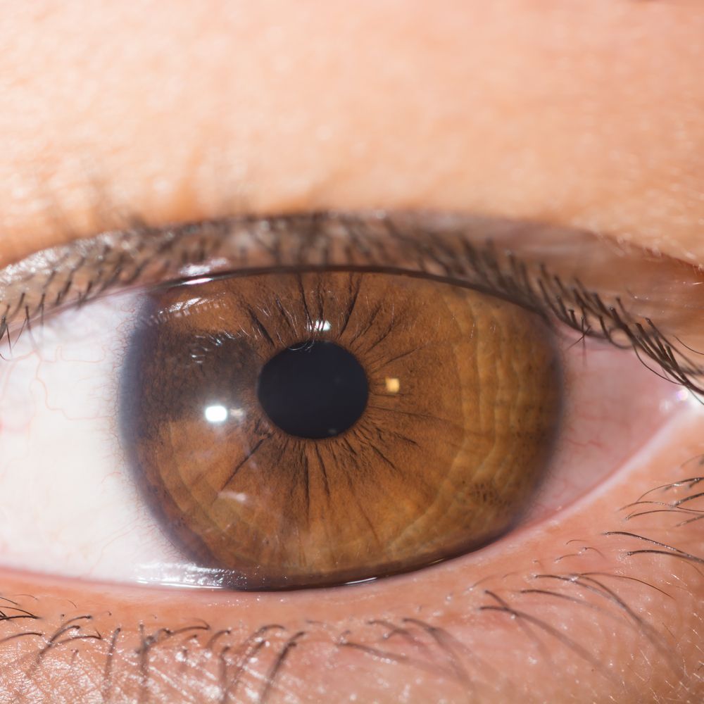
An eye with a freckle on the iris
Not only are nevi found on the front of the eye, but they can also be found further back in the eye where they are not visible to you. Inside the eye, choroidal nevi are very common findings. The choroid is a layer of tissue found beneath the retina (inner lining of the eye). Typically, choroidal nevi are found on routine eye examinations. A choroidal nevus will appear gray, yellow, or brown. It may be speckled or uniform in color. Many choroidal nevi of small or medium size are benign. The larger the nevus, the more concerned we become about choroidal melanoma.
Just like any freckle, or nevus, on the skin, there are many characteristics that are monitored to determine if it is benign or cancerous. For the eye, we follow the same ABCDE method to closely monitor these lesions. With any of these characteristics, the concern of cancer becomes elevated:
- A is for Asymmetry: If an imaginary line is drawn through the lesion, the sides would not be mirror images of one another.
- B is for Border: If the edges of the lesion are not crisp and defined but rather ragged, blurred or irregular.
- C is for Color: Variability in color meaning the lesion isn’t the same color all over. For choroidal nevi, that could mean orange pigment overlying the lesion.
- D is for diameter: For choroidal nevi, if the lesion is > 2.5mm.
- E is for Evolving: Changes or growth in nevus size, shape, and color.
Management of a nevus will vary depending on the characteristics it contains. Patients with no suspicious features will typically be monitored every year at their routine evaluations. With 2 or more suspicious features, a patient may be monitored more frequently. Other diagnostic tests may be performed to monitor a nevus including photography or OCT. It is important to have yearly routine eye exams with your doctor for early detection of any suspicious spots in the eye!
About the Author
Dr. Megan Fisher is an optometrist at Focal Pointe Eye Care in West Chester, Ohio, dedicated to providing comprehensive eye care for individuals and families.